


Past Researchers: Bjoern Andres
Collaborators (alphabetically): Davi Bock (Janelia Farm), Albert Cardona (Janelia Farm), Mitya Chlovskii (Janelia Farm), Winfried Denk (MPI Medical Research), Graham Knott (EPFL), Natalya Korogod (EPFL), Shawn Mikula (MPI Medical Research)
Research Focus
In order to better understand the structure and function of neural circuits, biologists today acquire huge amounts of three-dimensional image data with electron microscopy techniques such as SBFSEM and FIBSEM, with which an isotropic resolution of up to 5nm per voxel can be achieved.
Semi-automated and fully automated analysis of these datasets requires novel mathematical and algorithmical methods.
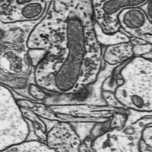
A typical two-dimensional slice (right) shows the membranes of the neurons, synapses, as well as intra-cellular structures such as mitochondria and vesicles.
Working at the interface of image processing, statistics and machine learning, we develop in close collaboration with our biological partners algorithms and tools to analyze these data sets.
Recovering the spatial structure of one or all neurons in the dataset, finding, counting and classifying synapses or other intra-cellular structures requires fast and accurate methods for interactive segmentation, automatic segmentation and synapse detection.
Interactive Segmentation

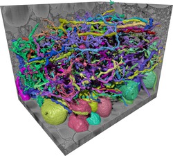
A fast and accurate method (Straehle et al, 2011) allows biologists to interactively recover the three-dimensional structure or segmentation of a single neuron of interest in a short amount of time. The method has been integrated into the Ilastik program (Website) with a convenient user-interface, such that only a few brush strokes are needed for an accurate segmentation.
References
- Seeded watershed cut uncertainty estimators for guided interactive segmentation
C. Straehle, U. Köthe, K. Briggman, W. Denk, F.A. Hamprecht
in: CVPR 2012. Proceedings, (2012), 765 - 772 [10.1109/CVPR.2012.6247747 | Technical Report] -
Carving: Scalable Interactive Segmentation of Neural Volume Electron Microscopy Images
C. N. Straehle, U. Köthe, G. Knott, F. A. Hamprecht
in: MICCAI 2011, Proceedings., Springer(2011) 6891, 603-660 [10.1007/978-3-642-23623-5_82 | Technical Report]
Automated Partitioning
In order to reconstruct the wiring diagram of a nervous system from an SBFSEM or FIBSEM volume image, accurate 3D image segmentation of all neurons contained in the data volume is required. As interactive segmentation would be much too costly, the ultimate goal is a completely automatic segmentation. However, established algorithms fail to provide the required accuracy.
A new method (Kroeger et al., 2012) for image processing goes beyond these algorithms by incorporating the non-local structure found in the volume image into the segmentation procedure. Moreover, segmentation criteria are not hand-crafted into the algorithm but are learned from a small subset of the data which was carefully traced by neuroscientists. Segmentation is finally cast into a global optimization problem which combines non-local image features with descriptors of the local image geometry.
References
-
Globally Optimal Closed-Surface Segmentation for Connectomics
T. Kroeger, B. Andres, K. L. Briggmann, W. Denk, N. Norogod, G. Knott, U. Köthe, F. A. Hamprecht
ECCV 2012. Proceedings, in press, (2012) [Technical Report] -
3D Segmentation of SBFSEM Images of Neuropil by a Graphical Model over Supervoxel Boundaries, in press
B. Andres, U. Köthe, T. Kröger, M. Helmstaedter, K.L. Briggman, W. Denk, F. A. Hamprecht
Medical Image Analysis, (2011) [10.1016/j.media.2011.11.004] -
Geometric Analysis of 3D Electron Microscopy Data
U. Köthe, B. Andres, T. Kröger, F. A. Hamprecht
in: Proceedings of Workshop on Discrete Geometry and Mathematical Morphology (WADGMM), (2010), 22-26 [Technical Report] -
How to Extract the Geometry and Topology from Very Large 3D Segmentations
B. Andres, U. Köthe, T. Kröger, F. A. Hamprecht
ArXiv e-prints, (2010) [URL | Technical Report] -
Segmentation of SBFSEM Volume Data of Neural Tissue by Hierarchical Classification
B. Andres, U. Köthe, M. Helmstaedter, W. Denk, F. A. Hamprecht
in: Pattern Recognition. 30th DAGM Symposium Munich, Germany, June 10-13, 2008. Proceedings, Springer(2008) 5096, 142-152 [10.1007/978-3-540-69321-5_15 | Technical Report]
Synapse detection
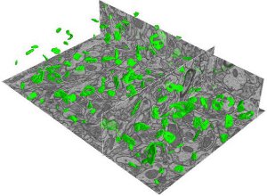
The chemical synapse is the predominant means by which information is transferred and stored in the central nervous system. Analysis of synapse size, shape and distribution contributes essential information to the understanding of neural circuitry, its function and its plasticity.
We propose a method (Kreshuk et al., 2011) which can automatically detect and segment synapses in a volume of neural tissue from a handful of example synapses outlined by a neurobiologist. Building on top of the classification capabilities in Ilastik, the algorithm learns a classifier with carefully selected 3D voxel features. On a quantitative validation data set with manual annotations by three independent experts the error rate of the algorithm was found to be comparable to that of the experts (0.92 recall at 0.89 precision).
We are currently working on the extension of the algorithm to data with anisotropic resolution (ssTEM data).
References
- Automated Detection and Segmentation of Synaptic Contacts in Nearly Isotropic Serial Electron Microscopy Images
A. Kreshuk, C. N. Straehle, C. Sommer, U. Köthe, M. Cantoni, G. Knott, F. A. Hamprecht
PLoS ONE, (2011) 6 (10)[10.1371/journal.pone.0024899] - Automated Segmentation of Synapses in 3D EM Data
A. Kreshuk, C. Straehle, C. Sommer, U. Köthe, G. Knott, F. A. Hamprecht
in: Eighth IEEE International Symposium on Biomedical Imaging (ISBI). Proceedings, (2011), 220-223 [10.1109/ISBI.2011.5872392 | Technical Report]